Welcome to Laser Pointer Forums - discuss green laser pointers, blue laser pointers, and all types of lasers

LPF Donation via Stripe | LPF Donation - Other Methods

Links below open in new window

ArcticMyst Security by Avery

putting together a gamma spectrometer (using soft MCA)

Joined
Jul 4, 2008
Messages
2,499
Points
113
Ahh I gotcha now Paul. I had a feeling you weren't wrong in what you were saying but you were saying it incorrectly, thus giving the wrong impression. No worries.

As with S_L I've never heard of that term (radiation toxicity) being used in the field (I don't have as much official experience as S_L but I did rad first-responder and some basic rad tech's rad safety courses). Even the term radiation poisoning is frowned upon; the term radiation sickness is what's used.

My apologies too Paul.... not trying to sound dick-ish. I had the CNRC regulations pounded into me doing the RAD-course.
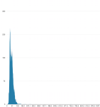
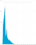
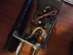
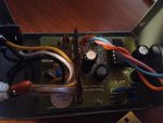
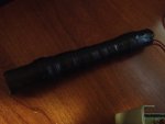
Anyways... I have just built a new scintillation detector with a new mini-CsI/Na crystal array. It's made in Russia. It's given so far very good results with low energy Gamma. pics are coming soon. The 59.8KeV (60KeV) peak is very distinct as well as the compton scatter and the lower energy X-ray(s).
I was testing it using several samples of the isotope. 0.9uCi, 5uCi, 50uCi and a 160uCi sample. The higher uCi sample caused the detector to oversaturate with the sheer counts. Over 4000cps @ 160uCi.. the 0.9uCi-5uCi samples produced a near perfect result over a 5 sec period.

I am using the GammaGrapher nano firmware on a broken DSO nano 1.7v.
It works but the software is full of issues. I am current just doing soundcard spectroscopy with it to get some useable data. so far that has worked better.
 
Last edited:


Joined
Sep 20, 2013
Messages
17,403
Points
113
SL, I'm looking forward to your spectrum pictures. I hope you have great luck with this project. ;)
 
Joined
Jul 4, 2008
Messages
2,499
Points
113
how's the process?

It's going. I've been having some medical tests done the past 2 or so weeks and on top mid-term for a Redseal course that I am taking.

Sorry for the delay.


I've attached the spectrum sample from a 10uCi Am241 source.
the 60.1KeV line is the largest peak, followed by the compton scatter to the right. The 26.5KeV line is to the left. It is barely visible as tiny spike.

The detector is a very tiny CsI/Na crystal hooked up to a 192Khz sound card and HV source with a pulse amplifier.
The result is this spectrum.

I suspect we are also seeing a clouding by Neptunium 237 in the sample as well. I suspect those are the peaks at 70KeV ?
More coming soon.
 

Attachments

  • Screen Shot 2016-02-24 at 11.32.57 PM.png
    Screen Shot 2016-02-24 at 11.32.57 PM.png
    47.9 KB · Views: 119
Joined
Jul 4, 2008
Messages
2,499
Points
113
A second Gamma spectrum from a very hot NORM sample. This sample is of a 80g or so sample of pure Uraninite from a place in British Columbia. The location is undisclosed. Note the huge 78KeV Pb X-ray peak and other peaks.
These are Uranium 238, Uranium 235, Pb214, Pb212, Bi214.... etc.. All of these are perfectly normal to see in a sample of Uranium mineralization.
To give you an idea how hot this sample is. Using a calibrated survey meter with a gamma only probe I measure 59,800CPM or approx 996CPS. The conversion of the probe is ~3600:1. I can get to 2.5KCPS on my scintillation detector easily as it is over 100x more sensitive to the lower energy Pb Xrays and other Uranium decay series gammas.

Yes, there are some strong U235 signature lines in this ore which is why some years ago the area was slated to be turned into a Uranium mine by the B.C. Government.
 

Attachments

  • Screen Shot 2016-02-25 at 8.10.09 PM.png
    Screen Shot 2016-02-25 at 8.10.09 PM.png
    52.5 KB · Views: 69
Joined
Jul 4, 2008
Messages
2,499
Points
113
This is a picture of the PMT driver. It is based on a Theremino PMT adapter which includes a HV low ripple, very low current source , a charge amplifier and a basic signal processor. HV starts at 700Vdc and tops at 1.5Kvdc.

The Scintillation detector is a very tiny setup that includes a CsI/Na.. these are very uncommon but work well. The crystal was provided to me from a contact in Russia. His crystals are very cheap and of excellent quality.
Tube was from IRad labs out of Florida it's a tiny 19mm dia tube,
called a Hamamatsu R1450. This is a perfect fit for mini-NaI/Tl, CsI/Tl, CsI/Na and PreludeTM420.

Anyways...
 

Attachments

  • IMG_3233.jpg
    IMG_3233.jpg
    135.4 KB · Views: 81
  • IMG_3234.jpg
    IMG_3234.jpg
    127.9 KB · Views: 86
  • IMG_3235.jpg
    IMG_3235.jpg
    117.8 KB · Views: 78
Joined
Sep 20, 2013
Messages
17,403
Points
113
SL, glad to see your spectrum and set up for your new scintillation detector, PMT and processor. I don't have a text containing sample spectra, so I'll have to get it somewhere else to analyze your results, but they look intriguing. :)
 
Joined
Jul 4, 2008
Messages
2,499
Points
113
SL, glad to see your spectrum and set up for your new scintillation detector, PMT and processor. I don't have a text containing sample spectra, so I'll have to get it somewhere else to analyze your results, but they look intriguing. :)

You'll have to have a look at other CsI/Na scintillation detector spectra. NaI/Tl is very distinct and has a 15-19% or so better resolving ability. I went with CsI/Na as it is less hydroscopic and has a higher density for resolving higher energy gammas. Those are 2 benefits I went after.
 
Joined
Sep 20, 2013
Messages
17,403
Points
113
You'll have to have a look at other CsI/Na scintillation detector spectra. NaI/Tl is very distinct and has a 15-19% or so better resolving ability. I went with CsI/Na as it is less hydroscopic and has a higher density for resolving higher energy gammas. Those are 2 benefits I went after.

I think you mean hygroscopic. That is the ability to absorb moisture. I know what you mean about the detector, though.
 
Joined
Jul 4, 2008
Messages
2,499
Points
113
Yes, that was a typo. Indeed Hygroscopic is the word intended here.
Also, CsI/Na is more durable to shock. It handles better.

I have compaired my spectra to my 1.25" dia NaI/Tl... It looks quite similar.

The real test will be with a test source of Cs137. I'll be looking for clean peaks at 32KeV (X-ray) and the high energy gamma peak at 662KeV.
If anyone has such a source, second hand I'd be happy to give it a new home :)

Looking for a Cs137 source at 1.0-5.0uCi. If you want to do a trade for a 50uCi Am241 button I'd be up for that too. :evil:
 
Last edited:
Joined
Sep 20, 2013
Messages
17,403
Points
113
A second Gamma spectrum from a very hot NORM sample. This sample is of a 80g or so sample of pure Uraninite from a place in British Columbia. The location is undisclosed. Note the huge 78KeV Pb X-ray peak and other peaks.
These are Uranium 238, Uranium 235, Pb214, Pb212, Bi214.... etc.. All of these are perfectly normal to see in a sample of Uranium mineralization.
To give you an idea how hot this sample is. Using a calibrated survey meter with a gamma only probe I measure 59,800CPM or approx 996CPS. The conversion of the probe is ~3600:1. I can get to 2.5KCPS on my scintillation detector easily as it is over 100x more sensitive to the lower energy Pb Xrays and other Uranium decay series gammas.

Yes, there are some strong U235 signature lines in this ore which is why some years ago the area was slated to be turned into a Uranium mine by the B.C. Government.

This has been bothering me for awhile because you mention the energy peaks from the U238 as Pb X-rays, which aren't what I have been accustomed to. All decay spectra are events that are alpha, beta or gamma particles and never an X-ray. But, some of the gamma peaks are in the energies of X-rays as they overlap gamma rays. From my text "Physics For Scientists and Engineers" X-Ray wavelengths are from 10^-8 m to 10^-13 m, while Gamma Ray wavelengths are 10^-10 m to 10^-14 m. clearly there's some overlap. So, I'm guessing what you are calling Pb X-rays are in fact gamma radiation. If, not, please explain. :thanks:
 
Joined
Jul 4, 2008
Messages
2,499
Points
113
This has been bothering me for awhile because you mention the energy peaks from the U238 as Pb X-rays, which aren't what I have been accustomed to. All decay spectra are events that are alpha, beta or gamma particles and never an X-ray. But, some of the gamma peaks are in the energies of X-rays as they overlap gamma rays. From my text "Physics For Scientists and Engineers" X-Ray wavelengths are from 10^-8 m to 10^-13 m, while Gamma Ray wavelengths are 10^-10 m to 10^-14 m. clearly there's some overlap. So, I'm guessing what you are calling Pb X-rays are in fact gamma radiation. If, not, please explain. :thanks:

X-rays and gamma rays overlap. yes..


I refer to Cs137 for example as having a 32KeV Xray and a 662KeV Gamma ray emission. It is due to their energy levels not their wavelength.
Technically you'd be correct in suggesting that because this emission is of "nuclear" origin. Under current nomenclature that is pretty standard thinking.
Regardless lower end emission being 32KeV it is still a gamma-ray HOWEVER....
There is quite an array of nomenclature on this x-ray vs gamma ray naming issue and more recent discoveries have challenged this notion. Take lightning for example.
It emits a gamma photon of only 10-20KeV.
 
Last edited:
Joined
Sep 20, 2013
Messages
17,403
Points
113
Hey, Seoul_lasers, haven't heard any more on your spectrometer lately. Was wondering if there is anything new?
 
Joined
Jul 4, 2008
Messages
2,499
Points
113
Hey, Seoul_lasers, haven't heard any more on your spectrometer lately. Was wondering if there is anything new?

I've moved into an apartment. I haven't had time to do any further experiments with the gamma spectrography driver.

Perhaps in the next several weeks or so I will get some time to conduct some experiments. :whistle:


Top